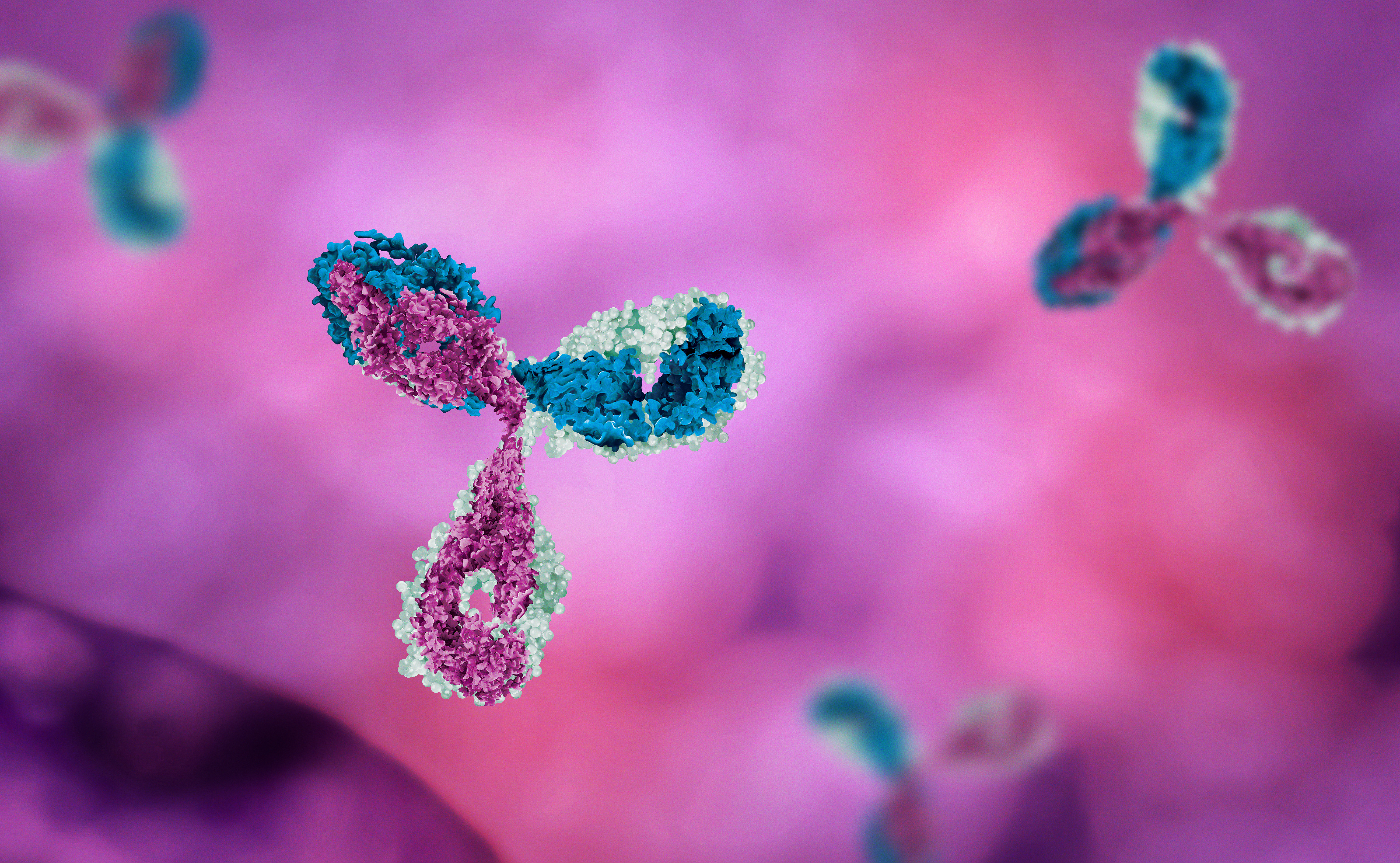
We have previously discussed the tremendous progress made towards the adoption of genomics medicine as standard of care in many clinical settings including cancer. In the field of oncology, it has become clear that personalised medicine powered by genomics knowledge (e.g. through liquid biopsies) is the future. In the field of prenatal testing, the use of genomics medicine is already part of the routine and we almost forget that it would have been science fiction not that long ago.
The cell free foetal DNA revolution
This story starts in the late 90’s, when Britney Spears was singing “Baby One More Time” and researchers discovered that foetal DNA could be detected in the blood of expectant mothers. For many readers, the former will feel like not that long ago: you can probably still sing that chorus from memory. Yet in that relatively short amount of time, the discovery of cell-free foetal DNA (cfDNA – the technical name for those fragments of foetal DNA circulating in maternal blood) changed the face of prenatal diagnosis. Indeed, if we can identify genomic sequences that belong to the foetus amongst the DNA present in the maternal sample, we can look at the genome of the foetus with a simple blood test. For example, we can identify the sex of the foetus (by detecting fragments of DNA derived from the sex chromosomes), detect chromosomal abnormalities (e.g. aneuploidies such as trisomy 21 causing Down Syndrome, by quantifying fragments derived from e.g. chromosome 21) and other genetic disorders (e.g. cystic fibrosis, by targeted detection of fragments derived from the CFTR gene), perform a paternity test (by detecting polymorphic loci that discriminate between the mother and alleged father), etc.
Compared to the main options that were available at the time to do many of these things (an amniocentesis or chorionic villus sampling (CVS), which are procedures where a sample of amniotic fluid or placental tissue, respectively, is obtained and tested for various genetic disorders), cffDNA based tests are revolutionary for two main reasons. Firstly, a blood draw is significantly less invasive than an amniotic fluid or CVS biopsy, and does not carry the same (small) risk of miscarriage. Secondly, the cffDNA can be detected in blood samples as early as the 7th week of gestation. By contrast, amniocentesis is typically performed around weeks 15-20 and CVS around weeks 10-12. These few weeks can make a world of difference to expecting parents.
The clinical reality
Since the 1990’s, many companies have developed diagnostic tests based on the detection of cffDNA. These include Roche’s Harmony®, Natera’s Panorama, Progenity’s innatal®, etc.
From a clinical perspective, cffDNA based tests are broadly categorised as non-invasive prenatal tests (NIPT, also referred to as screening) and non-invasive prenatal diagnosis (NIPD). The former are not tailored to the patient and provide as an output a risk of one or more conditions (typically limited to aneuploidies) and a sex determination - sex is also a numbers (of chromosomes) game. When a chromosomal aneuploidy is predicted by NIPT as being likely, a confirmatory (invasive) test will be offered to the patient. NIPD refers to a category of tests that is tailored to detect specific conditions (typically single gene disorders such as cystic fibrosis). This can be tailored to a patient, for example based on the patients’ family history. These tests are not necessarily followed by a confirmatory test.
Since 2018, NIPT has been used by the NHS as a first stage test, in combination with the first trimester ultrasound. Only if NIPT returns a positive test will parents be offered an amniocentesis. In this context, the level of certainty associated with the risk quantification is crucial. A lot of effort has gone into improving the sensitivity and specificity of NIPTs in the last couple of decades, both at the assay level and in the data processing steps.
Finding the needle in the haystack
One of the main challenges associated with these tests is that the cffDNA represents a small proportion of the DNA in the maternal plasma (referred to “foetal fraction”, this is typically around 5-15%). To put it simply, this means that we need to detect a small signal (cffDNA) through a lot of noise (maternal DNA – where the “noise” is 50% similar to the signal). This is further complicated by the fact that the amount of cffDNA in a sample changes from pregnancy to pregnancy, and during pregnancy. When it comes to detecting aneuploidies in particular, this is extremely important because we are effectively looking for an excess or under-representation of a particular foetal chromosome, compared to the expected (diploid) situation.
Over the last two decades, a lot of work has gone into improving our ability to (a) isolate and/or detect cffDNA (for example, based on the fragments size, their location in the genome, etc.), and (b) estimate the foetal fraction in a sample. One way to do this is to look for polymorphic loci that can differentiate between the mother and the foetus, and quantify those. This requires information about the genotype of the mother and foetus and a detecting technology that can discriminate between alleles at those informative positions (e.g. sequencing or specifically designed SNP microarrays). Another way to do this is to look at the size distribution of DNA fragments in the sample, as foetal fragments are typically shorter than maternal ones. Again, this typically requires massively parallel sequencing (which is still significantly more expensive than array-based methods). All of these approaches rely on custom bioinformatics pipelines, and so does the quantification of risk of abnormality in view of the determined foetal fraction (as previously mentioned, bioinformatics is behind much more of modern biology than you may think).
While there is still room for progress to be made in this field, the development and adoption of NIPT/NIPD solutions over the last few decades is an undeniable proof of the potential of data-driven genomics medicine to improve patients’ life.
Camille is a Partner and Patent Attorney at Mewburn Ellis. She does patent work in the life sciences sector, with a particular focus on bioinformatics/computational biology, precision medicine, medical devices and bioengineering. Camille has a PhD from the University of Cambridge and the EMBL-European Bioinformatics Institute. Her PhD research focused on the combined analysis of various sources of high-content data to reverse engineer healthy and diseased cellular signalling networks, and the effects of drugs on these networks. Prior to that, she completed a Master’s degree in Bioengineering at the University of Brussels and a Masters in Computational Biology at the University of Cambridge.
Email: camille.terfve@mewburn.com
Sign up to our newsletter: Forward - news, insights and features
Our people
Our IP specialists work at all stage of the IP life cycle and provide strategic advice about patent, trade mark and registered designs, as well as any IP-related disputes and legal and commercial requirements.
Our peopleContact Us
We have an easily-accessible office in central London, as well as a number of regional offices throughout the UK and an office in Munich, Germany. We’d love to hear from you, so please get in touch.
Get in touch

